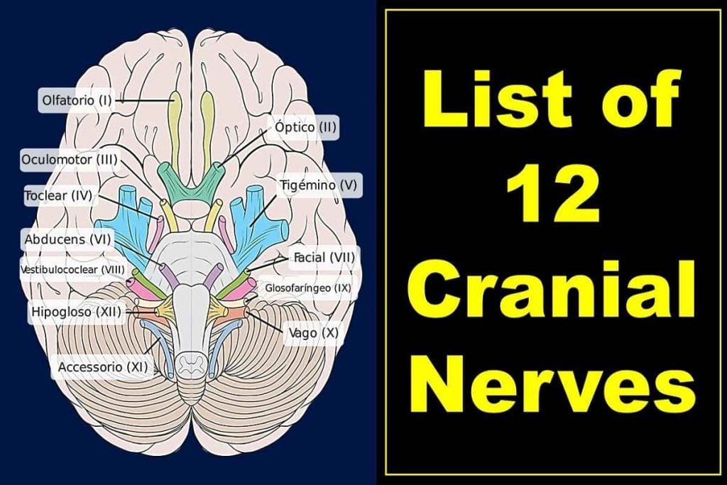
In this article, we will answer the question “What are the 12 cranial nerves?” We will also talk about the origin, anatomy, list of names in orders, and functions of 12 cranial nerves.
What are Cranial Nerves?
Cranial nerves are the bundle of paired nerves that connect your brain with your body. These nerves divide and send signals to different parts of your body, specifically to your brain, face, neck, and torso. They help you taste, smell and hear, as well as make facial expressions, blink your eyes and move your tongue.
The cranial nerves are a key part of the central nervous system in the body.

Functions of Cranial Nerves
Each cranial nerve has a different function related to movement or sense (sensory, motor, or both functions).
- Sensory neurons are the building blocks of sensory or afferent cranial nerves. A person may be able to hear, smell, or see because of these sensory neurons.
- Motor or efferent cranial nerves are made of motor neurons and they form the basis of the neck and head movements.
Recommended Article
Dirty, Funny Cranial Nerves Mnemonic to Memorize 12 Cranial Nerves
Origin of the 12 Cranial Nerves
Among 12 cranial nerves, the olfactory nerve (CN I) and optic nerve (CN II) are the only cranial nerves that originate from the cerebrum. The remaining cranial nerves III-XII emerge from the different parts of the brain stem, either medulla, pons, midbrain, or a junction between them.
The names of the cranial nerves are numerically identified in roman numerals. They are also related to their function and include I-XII.
What is the Largest and Longest Cranial Nerve?
The longest cranial nerve is the cranial nerve XI or vagus nerve. It is both sensory and motor in nature and runs through many parts of the body. It is present on the tongue, throat, heart, and digestive tract. Similarly, the trigeminal nerve (CNV), which supplies the eye and part of the face is the largest cranial nerve.
How many cranial nerves are there in a human body?
There are 12 pairs of cranial nerves. Each nerve of the pair innervates one side of the brain and body. For instance, the human body has a pair of optic nerves: one nerve of the optic nerve pair supplies the right eye, whereas another optic nerve of the pair innervates the left eye.
List of 12 cranial nerves in order
On the basis of the location of their exit site in the cerebrum (anterior to posterior) or origin site in the brainstem, cranial nerves are numbered in Roman numerals I to XII (superior to inferior origin location).
- Olfactory (CNI)
- Optic (CNII)
- Oculomotor (CNIII)
- Trochlear (CNIV)
- Trigeminal (CNV)
- Abducens (CNVI)
- Facial (CNVII)
- Vestibulocochlear (CNVIII)
- Glossopharyngeal (CNIX)
- Vagus (CNX)
- Accessory or Spinal Accessory (CNXI)
- Hypoglossal (CNXII)
Detail Explanation of Twelve Cranial Nerves
All of the 12 cranial nerves are unique in their pathway and functions. Here, you will know about individual cranial nerves.
Olfactory Nerve
The olfactory nerve transmits information to the brain about the smell.
When a person inhales fragrances, olfactory receptors send raw impulses to the cranial cavity. These impulses then travel to the olfactory bulb, where they are processed and turned into messages that our brains can better digest.
These specialized neurons and nerve fibers of the olfactory nerve meet with other nerves, which then pass into the olfactory tract.
The olfactory tract of the nose then travels to areas in the brain that deal with memory and notation of different smells.
Optic Nerve
In your eye, the optic nerve receives visual information and sends that information to your brain so you can see.
When light hits your eyes, it is detected by a variety of receptors in your retina. Rods are the most common type and are sensitive to light. These receptors allow you to see well during the night time but not during daylight hours.
There are fewer cones than there are rods. They also have lower light sensitivity and focus more on color vision.
Your eyes take in information through rods and cones located at the back of your retina. This invokes a response in your optic nerve, which transmits information to the occipital lobe, part of the brain that sits just behind your eyes. The place where these two optic nerves meet is called the optic chiasm, and it’s where fibers from both halves merge.
Through each optic nerve, the information eventually reaches your brain’s visual cortex. Here, the messages are processed and translated. The visual cortex is located in the back of our brains.
Oculomotor Nerve
The oculomotor nerve is a nerve that helps with eye movement.
Most of the muscles of the eyeball are innervated by the oculomotor nerve. This enables the smooth movement of the eyeball.
The oculomotor also plays a vital role in the involuntary functions of the eye.
The sphincter pupillae muscle regulates the amount of light your eyes get in. It is also the muscle that controls the size of an eye’s pupil.
The ciliary muscles help you to adjust eye focus by changing eye lens thickness. They work when you look at either near or far objects, allowing the lenses in your eyes to change automatically.
Trochlear Nerve
The trochlear nerve is one of the main nerves that control your superior oblique muscle. This muscle helps control your eye movements: out, down, and inward eye movement.
The trochlear nerve runs from back to front in your brain. It passes through the midbrain and reaches the eyes, which stimulates the superior oblique muscle.
Trigeminal Nerve
One of the most important nerves in your body, the trigeminal nerve is what helps control sensory functions and provides motor control. It is the largest cranial nerve.
The motor functions of the trigeminal nerve make it possible for people to chew and clench teeth, and they give sensation to the muscles in their ears.
Three divisions of the trigeminal nerves are ophthalmic, maxillary, and mandibular.
The intense pain and facial tics (trigeminal neuralgia) can happen with the disorder of the trigeminal nerve.
Abducens Nerve
The abducens nerve is a nerve that controls an independent muscle called the lateral rectus muscle. This muscle is usually responsible for outward eye movement and contraction.
The abducent nerve runs from your brainstem to your eye socket and controls one of the muscles (lateral rectus) for moving your eyes.
Facial Nerve
The facial nerve provides sensory and motor functions
- It helps moving facial and jaw muscles. Our facial muscles help us to determine emotion and the jaw muscle helps us form words.
- It also providing a sense of taste for most of your tongue
- The facial nerve supplies glands in your head or neck area, such as salivary glands and tear-producing glands
- It also communicates sensations from the outer parts of your ear
Your facial nerve starts in the pons area of your brainstem where it is also intertwined with a motor and sensory root. The two nerves eventually fuse together to form the facial nerve
Although the facial nerve may branch into smaller and larger fibers as it goes deeper, it does so only in order to stimulate muscles and glands or provide sensory information.
Vestibulocochlear Nerve
The vestibulocochlear nerve is known for providing the sense of hearing and balance in a person’s head.
The vestibulocochlear nerve contains two components: Vestibular and cochlear.
- The vestibular nerve, alongside input from other sensory systems and brain activity, can help you to keep your balance.
- Cochlear nerve, which is specialized to respond to sound and decide on frequency, allows the ear to pick up different signals. It plays an important role in our ability to get the most out of communication.
Glossopharyngeal Nerve
The glossopharyngeal nerve is divided into the motor and sensory functions. It also has autonomic functions, which include things like regulation of breathing and salivation.
- It sends sensory information from the back of your throat, sinuses, parts of your inner ear, and the back part of the tongue
- It also provides a sense of taste for the back part of your tongue
- The glossopharyngeal nerve also stimulates voluntary movement of a muscle in the back of your throat called the stylopharyngeus
The glossopharyngeal nerve originates in the medulla oblongata, a part of your brainstem. It then extends into your throat and neck region.
Vagus Nerve
The vagus nerve is very diverse. It has both sensory and motor functions, as well as parasympathetic actions.
The sensation information from the ear canal, parts of the throat, organs in the chest and trunk (heart and intestine) are communicated by the vagus nerve. It also controls the movement of the throat, the movement of the food in the digestive tract (peristalsis), and provides taste information at the root of the tongue.
The vagal nerve extends from your head to the abdomen and originates in the medulla part of your brainstem. It’s also the longest cranial nerve.
Accessory Nerve
The accessory nerve is one of the most important motor nerves in your body. It controls muscles that allow you to turn your head and neck into different positions, allowing you to bend over, among other movements
The accessory nerve is divided into two parts: spinal and cranial. The spinal part originates in the upper part of your spinal cord. The cranial portion starts in the medulla oblongata
Both of these sections come together for a short route. The spinal portion moves to innervate the neck muscles, whereas the cranial section goes along the vagus nerve.
Hypoglossal Nerve
Your hypoglossal nerve starts in your medulla and moves down into your jaw, where it activates most of the muscle in your tongue. It is the twelfth cranial nerve and its disorder leads to tongue paralysis.
Common Symptoms and Signs of Cranial Nerve Disorders
Your cranial nerves cannot always work as they should, which can lead to different problems. Depending on which cranial nerve is affected, you can have several disorders. The disorder of the cranial nerve affects your vision, taste, smell, facial expressions, body balance, hearing, and swallowing.
When Should You See the Doctor?
If you experience any of the following signs and symptoms, immediately call your doctor.
- pain, numbness, or drooping in one or both side of your face
- imbalance, weakness, or paralysis of any part of the body
- sudden droopy eyelid, squint, or partial/complete vision loss
- slurred speech
- loss of taste and smell
Takeaway
The cranial nerves in the human body send and receive signals from your face, neck, and torso. These nerves also play a role in movement and sensation.
If you experience a condition or injury that affects cranial nerves, it can result in problems with your sense of taste, smell, or vision.
If you’re experiencing difficulty maintaining normal facial expressions, such as constant drooping eyelids, eye squinting, numbness of the face, or others, it could be due to having a cranial nerve disorder.
Eating nutritious foods will help keep your whole body healthy, including cranial nerves. Also, consider adding a daily physical workout routine and managing any medical conditions for the normal functioning of the cranial nerves.


