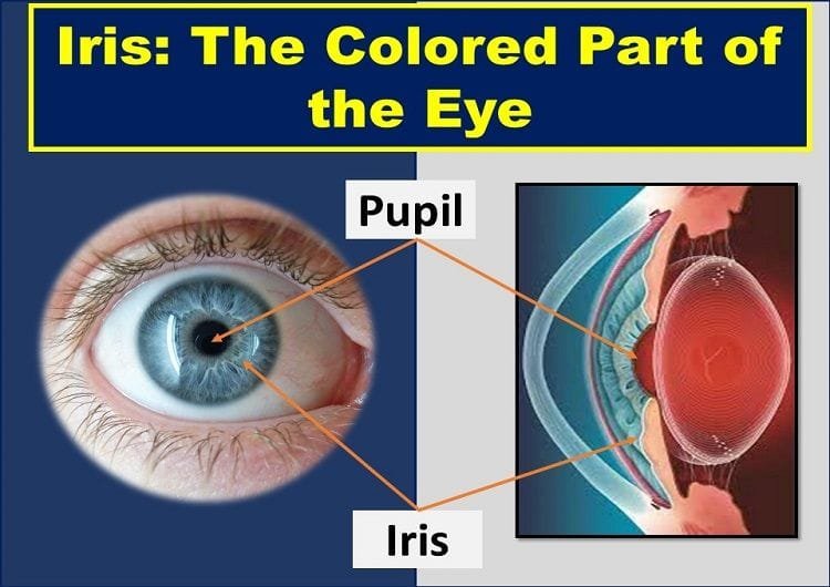
What gives color to the eye? What is the Iris of Eye? Today’s topic is all about the colored part of the eye or iris: Definition, Function, Anatomy, and Blood Supply. Stay connected.
What is Iris of Eye? (Iris Definition)
Iris is defined as the flat, annular structure behind the cornea and in front of the human crystalline lens. The iris of the eye is responsible for controlling the size of the pupil, which determines the amount of light entering the eye.
It divides the eyeball into the anterior segment and the posterior segment. The color of the eye is determined by the Iris color, as it is the colored part of the eye.
The central aperture of the iris is known as the pupil. The location of the pupil in the iris is not exactly central, it’s slightly inferior and nasal to the center. The pupil size determines the amount of light entering the eye.
The normal size (diameter) of the pupil is 3-4 mm. It varies based on the light condition in which the eye is exposed. The size of the pupil can also be altered pharmacologically.
Plural of Iris
The term “iris” was originated from a Greek word. In Greek mythology, the iris is the name of the Greek Goddess of the rainbow. The plural of the iris or the colored part of the eye is irises, irides, or just iris.
- Irises,
- Irides, or
- Iris

The Function of Iris or Colored Part of the Eye
Giving characteristic color to the eye is one of the functions of the iris. In addition to this, the major function of the iris is to act as a diaphragm of the eye (like a camera diaphragm) to control the size of the pupil (the central dark, round opening of the eye).
The dilator muscle of the iris (the dilator pupillae) increases the pupil size in the dark, whereas the constrictor muscle (the sphincter pupillae) constricts the pupil and limits the amount of light entering into the eye in the bright light.
This involuntary action of the iris (controlled by the brain) aids in clear and comfortable vision because too much or too little light can hamper vision.
Embryology or Development of the Iris of Eye
The development of the colored part of the eye, the iris starts from the optic cup. The anterior extension of the optic cup gives rise to the optic cup.
Both the anterior and the posterior epithelium are derived from the double-layered optic cup. Neuroectodermal cells of the optic cup are the sources of the muscles of the iris. Similarly, the iris stroma is formed from neural crest cells.
The important characteristic feature of the iris, the eye color, is dependent on the genetic character of the human.
Coloboma of the iris is the result of the failure of fusion of choroid fissure. The cleft on the inferior side gives a “keyhole” appearance. In a persistent pupillary membrane, the pupil is partially covered by the iris strands.
Likewise, Congenital aniridia is a complete absence of iris due to lack of development of optic cup.
Iris color or Eye Color
The eye color or the iris color is primarily determined by the amounts of melanocytes present in the stroma and anterior border layer of the iris.
The brown eye or the brown color of the iris is due to the abundant quantity of the melanocytes, whereas the paucity of the melanocytes results in the blue eye color.
In blue eyes, the longer wavelengths of light are absorbed, and the short wavelengths in the blue region of the spectrum are reflected or scattered, giving the blue appearance.
The iris color in albino is pale in color due to lack of pigmented melanocytes. The pinkish color of the iris is due to the transillumination of the iris by the light reflected from the retina.
The eye color or the iris color is inherited genetically. The brown color of the eye is inherited as a dominant trait, and the blue color is inherited recessively.
In some people, the iris has a different color between two eyes, or within the same eye (heterochromia).
Iris Anatomy
Iris Dimension
The colored part of the eye has an average diameter of 10-11 mm. It has a minimum thickness at the iris root, where it is about 0.5 mm thick.
It is the point at which the iris joins the ciliary body. Being the thinnest part of the iris, maximum damage occurs at this part of the iris during blunt trauma or injury to the eye.
The iris is thickest at the collarette. It is about 1.5 mm away from the pupillary margin.
Macroscopic Appearance of the Iris of Eye
Anterior Surface of the iris
The collarette or iris frill divides the anterior surface of the iris into a ciliary zone (posterior region) and the pupillary zone (anterior region).
Ciliary Zone
It is the part of the iris from the collarette to the iris root. It has radial streaks appearance because of the underlying radial blood vessels and crypts. The crypts are the depressions where the superficial layer of the iris is missing.
They are arranged in two rows: peripheral row near the iris root, central row near the collarette. Large crypts (Fuchs’s crypts) are found near the collarette.
Pupillary Zone
It is the part of the iris from the pupillary margin to the collarette. This region is relatively smooth and flat. There is a dark border at the pupillary margin, known as a pupillary frill or pupillary ruff.
The pupillary frill marks the anterior end of the pigmented epithelium at the posterior surface of the colored part of the eye.
Posterior Surface of the iris
The posterior surface of the iris is darker and smoother than the anterior surface. The darker appearance is due to the posterior pigment epithelium. There are several radial contraction folds and circular folds.
Microscopic Structure of Iris of Eye
Microscopically, there are four layers in the iris, namely from anterior to posterior: anterior limiting layer, iris stroma, anterior epithelial layer, and the posterior pigmented epithelial layer.
Anterior Limiting Layer
The anterior limiting layer is the anterior-most part of the iris that gives color to the eye, or its color determines the eye color. It consists of melanocytes and fibroblasts. This layer is missing in the area of the crypts.
In the blue eye or the blue iris, there is a thin anterior limiting layer and few pigment cells. But this layer is thick with densely pigmented cells in the brown iris or brown eye.
Iris Stroma
The large volume of the colored part of the eye is the iris stroma. It is made of a loosely arranged collagenous network and has dilator pupillae muscle, sphincter pupillae muscle, blood vessels, nerves, pigment cells, and other cells such as lymphocytes, macrophages, fibroblasts, and mast cells.
Among these cells, melanocytes, and clump cells are the pigmented cells, whereas fibroblasts, lymphocytes, macrophages, and mast cells are non-pigmented cells. Fibroblasts are found in the largest quantity in the iris stroma.
Muscles in the Iris Stroma
Two types of muscles found in the iris stroma are the sphincter pupillae muscle and the dilator pupillae muscle.
The sphincter pupillae muscle is 0.1 to 1.7 mm thick muscle which is around 1 mm in width and made of smooth muscle cells. As it is situated in the pupillary region of the iris stroma, the contraction of this muscle leads to constriction of the pupil or pupil miosis.
The sphincter pupillae muscle is supplied by the parasympathetic nervous system.
The dilator pupillae muscle is found in the ciliary zone of the iris stroma. The muscle fibers are arranged radially in this muscle hence its contraction pulls the pupillary portion of the iris towards the root, leading to pupil dilation or mydriasis.
The dilator pupillae muscle is innervated by the cervical sympathetic nerve system to dilate the pupil.
Anterior Epithelial Layer
The anterior epithelial layer is the anterior continuation of the posterior pigment epithelial layer and the pigmented epithelium of the ciliary body.
This layer is composed of two distinct portions: the anterior muscular basal portion (next to stroma), and the posterior apical portion (above posterior pigmented epithelium).
Posterior Pigmented Epithelial Layer
Posterior to the iris stroma lies the second layer of the pigmented epithelium, known as the posterior pigmented epithelium. It represents the anterior continuation of the non-pigmented epithelium of the ciliary body.
It becomes continuous with the anterior pigmented epithelium at the pupillary margin forming the pigmented frill.
The cells in this layer are more densely pigmented than the anterior pigment epithelium, hence the posterior surface of the iris is darker than the anterior surface.
This layer is made of rectangular or pyramidal cells that are joined to each other by maculae adherens and occludens. The basement membrane, where these cells rest, lies posteriorly.
Blood Supply to the Iris or Colored Part of the Eye
The uveal tract receives blood supply from three arteries: long posterior ciliary arteries, short posterior ciliary arteries, and anterior ciliary arteries.
Among these, long posterior ciliary arteries and anterior ciliary arteries are involved in the blood circulation of the iris. The long posterior ciliary artery is the branch of the ophthalmic artery.
There are seven anterior ciliary arteries that are derived from muscular branches of the ophthalmic artery. Two anterior ciliary arteries are supplied each from arteries of the recti muscles except the lateral rectus muscle.
Only one anterior ciliary artery is derived from the artery of the lateral rectus muscle.
Two long posterior ciliary arteries, one from the medial side and another from the lateral side, enter the sclera and reach the anterior uveal tract.
The superior and inferior branches of these long posterior ciliary arteries anastomose with each other and with the anterior ciliary arteries to form a circular blood vessel, the major arterial circle of the iris.
The branches of this major arterial circle of the iris enter the iris tissue and anastomose with each other to form a minor arterial circle to supply blood to the iris tissues and ciliary body.
Pupil and Iris Problems
The common problems of the colored part of the eye and pupil include:
Corectopia
Corectopia is defined as the abnormally placed pupil of the eye. The normal position of the pupil is the center (slightly nasal to the center) of the iris. So, eccentric placement of pupil is corectopia.
Corectopia is often associated with high myopia, ectopia lentis, among other conditions.
Polycoria
In polycoria, there is more than one pupil (or pupillary opening) in the eye. polycoria is extremely rare, and the cause of congenital polycoria is unknown.
In the case of true polycoria, the main pupil and other pupillary openings tend to be reactive to light and medication, whereas, in pseudopolycoria, there is passive constriction of the pupillary openings.
Dyscoria
The normal shape of the pupil of the eye is round. Any abnormality in the usual shape of the pupil is referred to as dyscoria.
Congenital Aniridia
The congenital absence of the iris, the colored part of the eye, is known as aniridia. True aniridia (complete absence of the iris) is an extremely rare medical condition.
In clinical aniridia, the peripheral rim of the colored part of the eye is present. Here, the ciliary processes and the lens zonules are visible. The clinical aniridia may be associated with glaucoma having angle anomalies.
Synechiae
The medical term ‘synechia’ refers to the condition in which the iris adheres to the front surface of the crystalline lens or the back surface of the cornea, the transparent, anterior most structure of the eye.
The common causes of the synechiae are trauma uveitis (iritis), among other causes. Synechiae should be treated as soon as possible as they can lead to angle-closure glaucoma by blocking the normal aqueous drainage pathway.
Persistent Pupillary Membrane (PPM)
Persistent pupillary membranes (PPMs) are the congenital remnants of the vascular sheath of the lens that are seen floating freely in the anterior chamber or attached to the anterior surface of the lens.
The pupillary membranes are the source of blood supply to the lens in the fetus.
Congenital Coloboma of the Iris
The medical term ‘coloboma’ means the absence of tissue. The iris coloboma is a congenital condition in which the portion (mostly inferior region) of iris tissue is missing, giving the cat-eye or keyhole appearance of the pupil.
They can be seen in association with other tissues of the uveal tract or independently.
The most common type of coloboma, typical coloboma, is found in the inferonasal region of the iris as a result of defective closure of embryonic fissure.
If coloboma is seen in other quadrants, it is known as atypical coloboma. Similarly, the complete coloboma might extent from the pupil to the optic nerve.
Iris Melanoma
Iris melanoma is the cancerous tissue mass in the colored part of the eye, the iris. It develops from the pigment cells or melanocytes present in the iris.
Iris melanomas are one of the common types of cancer in the eye, and if metastasis occurs, it can affect other parts of the body. Treatment of iris melanoma is effective if diagnosed at the early stage, but in severe cases, or in late stages, enucleation (removal of the affected eye) may be necessary.
Iris Nevus
Iris nevus (plural nevi) or freckle is a pigmental mass in the iris. It is a stable and harmless pigmentary mass like freckles on the skin. If the size of the nevus is getting larger, it might show a sign of malignant growth. Visit your eye doctor in such rare circumstances.
Anterior Uveitis (Iritis)
Iritis is a type of anterior uveitis, and it is the inflammation of the colored part of the eye. If the inflammation is not limited to the iris and if it affects the ciliary body along with the iris, it is known as iridocyclitis.
It is one of the vision-threatening conditions in the eye. The clinical signs and symptoms of the iritis include eye pain, redness, sensitivity to light (photophobia), and reduced vision (visual acuity).
YOU MAY ALSO LIKE
Diplopia Charting: Common Method of Double Vision Test
Hess Charting: Interpretation of Hess Screen Test
Blurry Vision In The Morning – Causes And Concerns
Wavy Squiggly Lines in Vision (Eye Floaters)
Bulbar Conjunctiva, Palpebral Conjunctiva, and Fornix of Eye
What is Conjunctiva of the Eye?
White Part of the Eye: Sclera Function, Definition & Anatomy
What is Cornea of Eye: Function, Definition, Anatomy, Layers
Cranial Nerves Mnemonic: How to Memorize Fast
Right & Left Eye Twitching Meaning
Sudden Blurry Vision in One Eye: Causes, and Treatment
References
- Wolff’s Anatomy of the Eye and Orbit by Anthony J. Bron, Ramesh C. Tripathi, Brenda J. Tripathi
- Comprehensive Ophthalmology Fourth Edition by A. K. Khurana


