All about Trochlea in the human body: Trochlea in Humerus, in Femur, in Superior Oblique Muscle in Eye, and many more. Stay connected.
Definition
In medicine, the trochlea is defined as an anatomical structure that resembles a pully or grooved structure. The term “trochlea” is derived from a Latin word that means “case or shear containing one or more pulleys, block”.
Pully-like anatomic structures are found in several locations within the living body: in the elbow joint, the knee joint, the eye orbit, and many others.
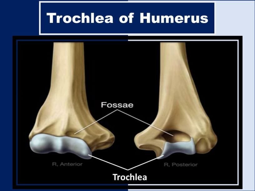
Trochleae are commonly seen in joint bones to provide an articular surface. The humeral trochlea’s articular surface of the medial condyle articulates with the trochlear notch of the ulna. It is an important component of the elbow hinge joint.
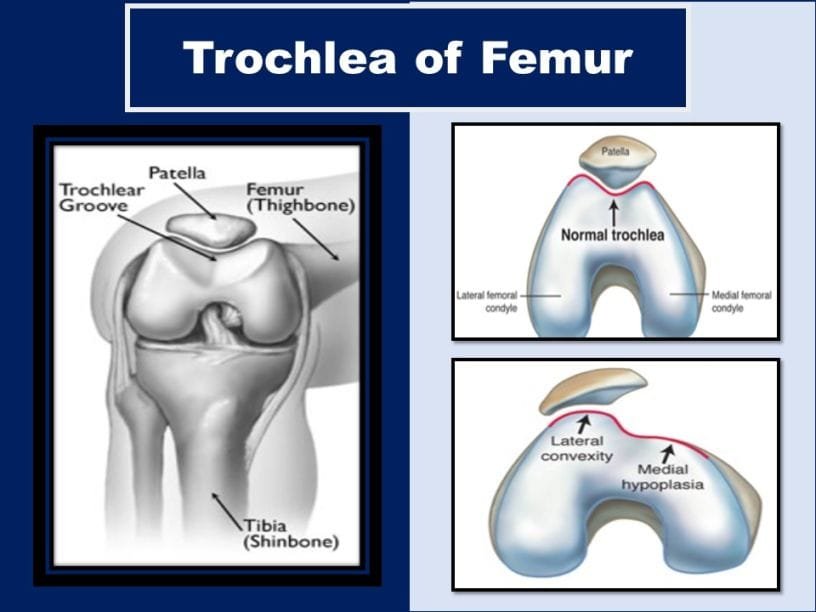
The knee hinge joint has a femoral trochlea to join the patella with the distal femur. Similarly, in the eye orbit, it provides a pathway or guides for superior oblique muscle for smooth eye movement.

Humeral Trochlea
In humans, the lower end of the humerus has two smooth articular surfaces, two depressions (fossae), and two projections (epicondyles). Two articular surfaces are the capitulum and trochlea.
The capitulum, located laterally, articulates with the radius whereas the humeral trochlea that is located medially articulates with the ulna.
The two fossae that form the part of the elbow joint are the olecranon fossa and the coronoid fossa. The olecranon fossa is situated behind and above the humeral trochlea whereas the coronoid fossa is found in the front and above the humeral trochlea.
These fossae receive projections from the ulna as the elbow is alternately straightened and flexed. Likewise, the epicondyles provide attachment for muscles which is necessary for movements of the forearm and fingers.

Function
The main function of the trochlea of the humerus is to provide a smooth surface that articulates with the notch. This structure in association with notch, fossae, and epicondyles provide smooth functioning of the elbow hinge joint (elbow flexion and extension). The ossification of this structure begins at 8-9 years of age.
Femoral Trochlea
The trochlea of the femur or thigh bone is a bony structure located on the femur’s distal end, and part of the patellofemoral articulation that also encompasses the anterior section of the intercondylar fossa.
It provides a channel-like groove or surface for the attachment of leg bones together. The femoral trochlea is the key component of the knee joint or the patellofemoral joint.
It consists of a groove bounded by the medial and lateral ridges. These ridges are asymmetrical in large animals and in horses, there is a protuberance on the proximal part of the medial ridge called the tubercle of the trochlea of the femur.
Normal trochlear bony anatomy contributes to knee joint stability. Acquired or congenital changes in the surface geometry of the joint can lead to various clinical disorders, such as chondromalacia patella, instability in the patella, and joint pain.

Function
Proper location of the trochlea of the femur is the most for smooth functioning of the knee cap or patella. Without the femoral trochlea, the patella is unable to maintain its normal position.
Knee problems and pain can arise if the knee cap slides away from the trochlea of the femur. Patella dislocation or subluxation is the condition in which the knee cap is displaced from its position.
Injuries or other knee malformations, such as excessive rotation of the femur (femoral torsion), can cause the knees to turn inwards toward each other and create a poor alignment of the knee inside the femoral trochlea, causing pain and dysfunction.
Trochlea of Talus
It is situated at the superior surface of the body of the talus. It provides a smooth surface for articulation with the tibia.
The trochlea is broader in front than behind, convex from before backward, slightly concave from side to side: in front, it is continuous with the upper surface of the neck of the bone.
Peroneal Trochlea
On the lateral surface of the calcaneus, in front of the tubercle or the attachment of the calcaneofibular ligament, it presents a narrow surface marked by two oblique grooves.
The grooves are separated by an elevated ridge, or tubercle, the fibular trochlea (trochlear process, peroneal tubercle, peroneal trochlea), which varies much in size in different bones.
The superior groove transmits the tendon of the Peroneus Brevis; the inferior groove, that of the Peroneus longus (called the Groove for the tendon of fibularis longus; Groove for the tendon of peroneus longus).
Trochlea of Superior Oblique Muscle in the Eye
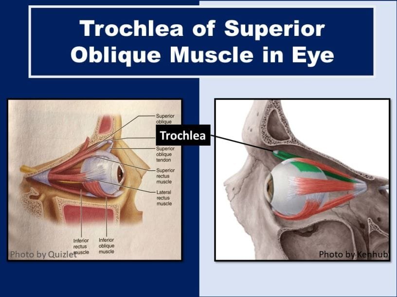
It is a pully-like anatomical structure found in the orbit. It provides the guide or pathway to the tendon of the superior oblique muscle. The trochlea of the eye is located on the superior nasal aspect of the frontal bone. Interestingly, it is the only cartilage noticed in the normal eye orbit.

Function
The major function of the trochlea in the eye is to maintain the eye movement action of the superior oblique muscle. The primary (main) action of the superior oblique muscle is intorsion (internal rotation), the secondary action is depression (primarily in the adducted position) and the tertiary action is abduction (lateral rotation).
Sources
- https://www.imaios.com/en/vet-Anatomy/Vet-Anatomical-Part/Trochlea-of-femur
- Botchu R, Obaid H, Rennie WJ. Correlation between trochlear dysplasia and the notch index . J Orthop Surg 2013;21:290-3.
- https://www.bartleby.com/107/
- Murshed KA, Cicekcibası AE, Ziylan T, Karabacakoglu A. Femoral sulcus angle measurements: Anatomical study of magnetic resonance images and dry bones. Turk J Med Sci 2004;34:154-169.
- Tecklenburg K, Dejour D, Hoser C, Fink C. Bony and cartilaginous anatomy of the patellofemoral joint . Knee Surg Sports Traumatol Arthrosc 2006 ;14:235-40.
YOU MAY ALSO LIKE
Beck’s Triad in Cardiology: Mnemonic, Treatment & More
Cushing’s Triad in ICP: Pathophysiology, Mnemonic, & More
Acronym for Cranial Nerves Mnemonic – Dirty, Funny Tricks
Spermatic Cord Contents Mnemonic – Dirty and Funny
Carpal Bones Mnemonic: Wrist Bones Names in Order
Essential Amino Acids Mnemonic: How to Memorize Fast!


