All About Lines in Eyes or Lines in Ophthalmology: Arlt’s Line, Hudson Stahli Line, Khodadoust Line, Stocker lines, Vogt’s and Haab’s Striae, and More.
Lines in Eyes or Lines in Ophthalmology gives you a comprehensive list of all the important streaks, striae, and lines observed in all structures of the eye; from the anterior-most cornea to the posterior retina and orbit wall. This important list of lines in eyes is equally useful for medical students, medical practitioners, paramedics, nurses, and researchers in the field of medicine.
The list can be a companion for medical students preparing for any type of entrance exams and university exams. Lines in Eyes (Ophthalmology) is a useful nursing guide for those who are participating in USMLE and other academic and professional exams. So, without further ado let’s jump into the topic.

Stocker Line
The Stocker line is a vertical, yellow-brownish line found along the advancing edge of the pterygium. It is formed by the deposition of hemosiderin (a form of iron) in the conjunctival and corneal epithelial layers. When seen in blue light (cobalt filter), the pigment of Stocker’s line looks dark due to a lack of fluorescence.
Stocker’s line is a punctate, subepithelial line (or lines) passing in front of the invasive apex of the pterygium.
The exact mechanism of iron deposition in the development of advancing pterygium and the formation of the Stoker line is still unknown, but the level of iron is seen in a significantly higher proportion in the pterygium tissue than in the normal conjunctival tissue.
According to Hiroto Obata, et. al., Stocker’s line is rarely observed in a small pterygium, and the occurrence of the Stoker line is related to the time duration from the onset of the pterygium in the eye.
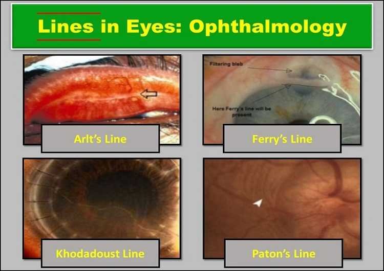
Arlt’s Line in Eye
Arlt’s line is a hallmark of trachoma, a severe infection of the eye caused by Chlamydia trachomatis. The infection leaves a thick band of scar tissue in the conjunctiva near the eyelid margin.
Arlt’s line in the eye is a horizontal line found at the junction of the anterior one-third and posterior two-thirds of the conjunctiva (sulcus subtarsalis). Arlt’s line (lines) runs parallel to the eyelid.
The Arlt’s line got its name after the famous Austrian Ophthalmologist Carl Ferdinand von Arlt.
Schwalbe’ Lines in Ophthalmology
Schwalbe’s line is a thin, white, or irregularly pigmented line found on the interior surface of the cornea of the eye. It represents the peripheral margin or outer limit of the corneal endothelium layer. More precisely, Schwalbe’s line represents the termination of the Descemet’s membrane of the cornea. The detail of the line can be observed through gonioscopy.
Sampaolesi Lines in Eyes
Sampaolesi line is a wavy line found anterior to Schwalbe’s line in pigment dispersion syndrome, pseudoexfoliation syndrome, trauma, or iris melanoma. It is the hyperpigmentation line seen through gonioscopy. Gonioscopy involves the placing of a mirrored lens on the patient’s cornea to examine the angle structures of the anterior chamber of the eye.
No treatment is necessary for the Sampaolesi line in the eye but it is vital to monitor for glaucomatous changes.
Hudson-Stahli Line in Ophthalmology
The Hudson-Stahli line is a line formed by iron deposition. It is located roughly on the border between the middle and lower thirds of the cornea (corneal epithelium). The superficial horizontal brown line was independently described by Hudson in 1911 and Stahli in 1918.
The Hudson-Stahli line is about o.5 mm thick and 1-2 mm long. This line is not caused by any ocular or systemic diseases. It is present normally in older people but seems to dissipate to some degree by the age of 70.
Some studies have shown that the Hudson-Stahli line is enhanced by chloroquine and hydroxychloroquine toxicity. The line formation is also dependent upon the rate of tear secretion.
Khodadoust Line in Eye
Also known as chronic focal transplant reaction, a Khodadoust line is a medical sign that indicates a complication of corneal graft surgery (corneal transplant). The line is named Khodadoust after the researcher of this line, Prof. Ali Asghar Khodadoust.
The medical condition involves the immunological rejection of a transplanted cornea which is not seen in internal organ transplant and rejection.
A Khodadoust line is made up of white blood cells (mononuclear cells) at the vascularized edge of the recently transplanted cornea.
Immediate immunosuppression treatment is necessary to prevent further damage because, without treatment, the line of WBC will move across and damage the endothelial cells within several days.
Ferry’s Lines in Eyes
Ferry’s lines in eyes are observed in the corneal epithelium at the edge of the filtering bleb in glaucoma. Filtering bled is a conjunctival blister resulted from glaucoma surgery to lower the intraocular pressure. Here, a flap of the sclera is created in the eyewall which allows the aqueous humor to drain out of the eye and underneath the conjunctiva.
Zentmeyer Line or Scheie’s Line
Zentmeyer line or Scheie’s line is defined as a peripheral pigmentation of the posterior lens capsule anterior to the junction between the anterior hyaloid face and the posterior lens capsule – ligamentum hyaloideo-capsulare of Wieger.
Zentmeyer’s lines in ophthalmology are the hallmarks of pigment dispersion syndrome.
Ehrlich-Turck Lines in Ophthalmology
The Ehrlich-Turck lines in eyes are seen on the endothelium of the cornea due to a linear deposition of keratic precipitates (KPs) in uveitis.
Paton’s Lines in Eyes
Paton’s lines in ophthalmology are the circumferential retinal folds formed in the peripapillary region due to papilledema. Papilledema is defined as optic nerve head edema secondary to increased intracranial pressure (ICP). The main cause of optic nerve head swelling is blockage of the axoplasm transport and the blockage occurs at the lamina cribrosa.
The optic nerve head can swell to the extent where it is extended forward into the vitreous as well as laterally. This lateral swelling causes the retina to buckle inward at the temporal aspect of the optic nerve head. The buckling is known as Paton’s lines or folds.
Rucker’s Lines in Eyes
Rucker’s line in eye is formed by the sclerosed vessels due to periphlebitis retinae. It is a hallmark of multiple sclerosis.
Schlagel’s Lines in Ophthalmology
Schlagel’s lines in eyes are the multiple yellow lines seen at the posterior pole and peripheral retina. The lines are arranged in clumps or linear streaks, commonly seen in multifocal choroiditis.
White Lines of Vogt
White lines of Vogt are the sheathed or sclerosed vessels found in Lattice degeneration. Lattice degeneration of the retina is a fairly common degenerative disease of the peripheral retina. It is characterized by the presence of lattice lines created by fibroid blood vessels.
Crisscrossing fine white lines of Vogt that account for the name lattice degeneration is present in roughly one-tenth of lesions and most likely represent hyalinized blood vessels.
Fingerprint Lines in Eyes
Fingerprint lines in ophthalmology are seen in map-dot fingerprint dystrophy of the cornea. Corneal map-dot fingerprint dystrophy is the most common corneal dystrophy.
The name is for the appearance of its characteristic slit-lamp findings. Other names of map-dot-fingerprint dystrophy are epithelial basement membrane dystrophy, anterior basement membrane dystrophy, and Cogan microcystic epithelial dystrophy.
LASIK Iron Lines in Eyes
After LASIK eye surgery for myopia, the central corneal curvature is flattened than before surgery. As a result, the tear film distribution is altered, allowing some pooling centrally. This pooling causes iron deposition in the central epithelium.
A similar effect can be seen after steeping of the cornea from the treatment of hyperopia. In the case of hyperopia, a Pseudo-Fleischer’s Ring (iron deposition) can be seen. These iron lines in the eyes are visually insignificant.
Waring Lines in Ophthalmology
Waring lines are common in eyes after radial keratotomy (RK). These lines are present in up to 80 percent of RK eyes. The Waring line is formed by the stellate corneal epithelial iron deposition.
Krukenberg’s Spindle
Krukenberg’s spindle is the pattern formed on the inner surface of the cornea by pigmented iris cells which are deposited due to the currents of the aqueous humor.
The phenomenon of formation and the sign of Krukunberg’s spindle was first described in 1899 by Friedrich Ernst Krukenberg (1871-1946), a German pathologist who specialized in Ophthalmology.
Siegrist Streaks
Siegrist streaks are hyper-pigmented flecks that are arranged in a linear fashion along the choroidal blood vessels. These are seen in hypertensive choroidopathy and fibrinoid necrosis.
Ohngren’s Lines in Eyes
Ohngren’s line is seen in orbit. An x-ray description of the line dated back to 1930. Ohngren’s line delineates the limits of resectability of maxillary sinus tumors. If the line is present in supero-posterior position, it is likely to invade orbit, ethmoids, and pterygopalatine fossa.
Vogt Striae
Vogt’s striae are vertical (rarely horizontal), whitish lines found in the deep/posterior stroma and Descemet’s membrane. It is common in patients with keratoconus. Vogt’s striae can be asymmetric depending on the degree of keratoconus in each eye.
Vogt’s striae are also known as Descemet’s membrane lines.
There is a positive correlation between the orientation of the lines with the steepest axis of the cornea. The formation of striae is related to mechanical stress forces on collagen lamellae radiating from the cone apex. The Vogt’s striae can temporarily disappear with external pressure to the globe.
Haab Striae
Haab’s striae or Descemet’s tears are horizontal breaks in the Descemet’s membrane and are associated with congenital glaucoma. It is named after Otto Haab.
The appearance of Haab’s striae is similar to posterior polymorphous dystrophy (PPMD). However, on histopathology, the edge of Haab’s striae are thickened, curled, with the area between the edge being smooth and thin. This helps differentiate Haab’s striae from PPMD.
Sources
- https://iovs.arvojournals.org/article.aspx?articleid=2561730
- https://www.whonamedit.com/synd.cfm/3947.html
- https://www.eyenews.uk.com/media/11348/eyeam15-trainees2.pdf
YOU MAY ALSO LIKE Rings in Eyes (Ophthalmology): Cholesterol, Blue, & More


