Your eye captures light. It turns that light into electrical signals. But those signals need a path to your brain. That path is the optic nerve. It is the cable that connects your eye to your brain. Without it, you would be blind, even if your eyes were perfectly healthy.
The optic nerve is the second cranial nerve, or CN II. It is made of more than one million tiny nerve fibers. These fibers come from the retina, the light-sensitive layer at the back of your eye. They bundle together and leave the back of your eye through a hole in the sclera, the white part.
The optic nerve travels from your eye to your brain. The two optic nerves, one from each eye, meet at a place called the optic chiasm. Here, some fibers cross over to the other side of the brain. This crossover helps your brain combine what both eyes see into a single, three-dimensional image.
The optic nerve has no pain fibers. That is why you cannot feel it. But it can be damaged by disease. Glaucoma, tumors, and inflammation can all hurt the optic nerve. Once damaged, it cannot heal.
Let’s explore the optic nerve, including its definition, function, and anatomy. We will learn how this vital cable brings vision to your brain.
What is Optic Nerve?
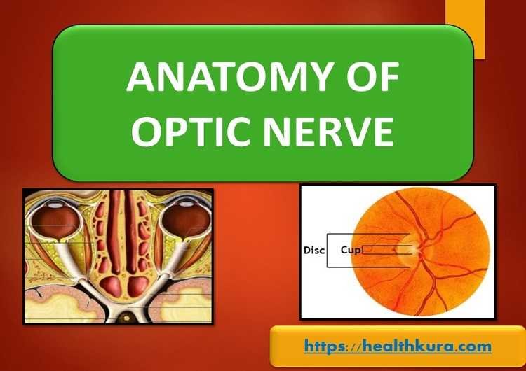
Also known as cranial nerve II or simply as CN II, the optic nerve is a paired cranial nerve that carries visual impulses from the innermost layer of the eye, retina to the brain. It establishes the connection between the eye and the brain.
These visual impulses, dispatched through the optic nerve to the brain, form the building blocks of the image of the object. The second cranial nerve is the only visible part of the brain (or its extension), and the optic nerve head (optic disk) can be easily viewed by using an ophthalmoscope.
The second cranial nerve is a part of the central nervous system as it is derived from the out-pouching of the diencephalon during embryogenesis. Being the cranial nerve, the CN II is covered with myelin produced by oligodendrocytes, rather than Schwann cells of the peripheral nervous system.
Like other cranial nerves, the optic nerve is ensheathed in all three meningeal layers (dura, arachnoid, and pia mater) rather than the epineurium, perineurium, and endoneurium found in peripheral nerves.
The CN II is formed by glial cells and more than 1 million nerve fibers which are axons of the retinal ganglion cells of the retina.
Optic Nerve Function
All sorts of visual information, such as the perception of brightness, contrast, color perception, are transmitted via the optic nerve. It also plays a role to conduct two important neurological reflexes, light reflex, and accommodation reflex. The light reflex is necessary for constriction and dilation of both pupils according to the amount of light shone into the eyes. Likewise, accommodation reflex facilitates the eye to adjust the lens thickness for clear near vision.
Examining the anatomical integrity and functions of the CN II, eye care professionals can determine the health status of the visual pathway and the areas nearby the visual pathway and visual cortex. For instance, the pituitary adenoma can be suspected from the abnormal functioning of the optic nerve. Likewise, increased intracranial pressure leads to papilledema, which can be easily examined.
Development
The optic nerve develops in the framework of the optic stalk. To form the CN II fibers, the fibers from the retinal nerve fiber layer grow into optic stalk by passing through the choroidal fissure. The glial system of the nerve is developed from the ectodermal cells of the walls of the optic stalk.
The fibrous septum develops from the vascular layer of mesenchyme at the third month of gestation. Similarly, CN II sheaths are developed from the mesenchyme layer similar to the meninges of other parts of the central nervous system. Myelination of nerve fibers starts from the brain and extends up to the lamina cribrosa just before birth. If myelination extends up to around the optic disc, it presents as congenital myelinated nerve fibers.
Anatomy of the optic nerve and visual pathway
The visual pathway starts from the innermost layer of the eyeball, retina and extends up to the cortical region of the brain consisting of the optic nerve, optic chiasma, optic tracts, lateral geniculate bodies, optic radiations, and the visual cortex.
Parts of the Optic nerve
About 47-50 mm long CN II is divided into 4 parts: intraocular (1 mm), intraorbital (30 mm), intracanalicular (6-9 mm), and intracranial (10 mm).
Optic chiasma
Optic chiasma is 8-12 mm flattened structure that lies over the tuberculum and diaphragm sellae. The optic nerve fibers from the nasal halves of the retina get decussated at the optic chiasma.
Optic tracts
The cylindrical nerve fiber bundles run posteriorly from the optic chiasma. It consists of nerve fibers from the nasal half of the retina of the opposite eye and the temporal half of the same eye. The optic tract end in the lateral geniculate body.
Lateral geniculate bodies
It is located at the posterior termination of the optic tract. It is formed by six layers of grey matter (neuron) alternating with white matter. The second-order neurons coming through the optic tract relay information to the lateral geniculate bodies.
Optic radiations
It is formed by the axons of third-order neurons of the visual pathway and extends from the lateral geniculate body to the visual cortex.
Visual cortex
The visual cortex is located on the occipital lobe, below and above the calcarine fissure. The subdivisions of the visual cortex are the visuosensory area (striate area 17) that receives optic radiation fibers, and the surrounding area (peristriate area 18 and parastriate area 19).
Blood supply of the visual pathway
Ophthalmic branches of the internal carotid artery, posterior ciliary arteries, and central retinal artery supply blood to different parts of the optic nerve. Most of the parts of the visual pathway are supplied by the pial network of blood vessels except the orbital part of the optic nerve. The pial network (pial plexus) is formed from different arteries.
The capillaries derived from the retinal arterioles supply the surface layer of the optic disc. The prelaminar region of the nerve head gets blood supply from the branches of the peripapillary choroid and some contributions from the vessels of the lamina cribrosa. Likewise, the posterior ciliary arteries and arterial circle of Zinn supply blood to the lamina cribrosa.
The retrolaminar part of the second cranial nerve gets blood supply from the centrifugal branches of the central retinal artery and branches from the pial plexus formed by branches from the choroidal arteries, circle of Zinn, central retinal artery, and ophthalmic artery.
Venous Drainage
The major vein involves in the venous drainage of the optic nerve is the central retinal vein. The pial venous system is also responsible for venous drainage to a lesser extent. Both systems drain into the ophthalmic venous system in the orbit and less commonly directly into the cavernous sinus.
Fibers
Visual afferent fibers are responsible for transmitting visual impulses from the retina to the lateral geniculate body of the thalamus. Likewise, pupillary afferent fibers regulate the pupillary light reflex. Efferent fibers travel to the retina but have an unknown function. Similarly, photostatic fibers are responsible for visual body reflexes.
Blood-brain barrier at the optic nerve
The non-fenestrated endothelial linings with tight junctions between the adjacent endothelial cells are present in the capillaries of the optic nerve head, the retina, and the central nervous system. These tight junctions act as the blood tissue barrier to the diffusion of small molecules across capillaries. But this junction is incomplete as a result of continuity between the extracellular spaces of the choroid and the prelaminar region of the optic disc. There is no blood tissue barrier to diffusion across the highly fenestrated capillaries of the choroid.
What are the signs of optic nerve dysfunction?
The following signs are more common in the defect of optic nerve function.
- Reduced visual acuity (VA)
- Afferent pupillary defects
- Visual field defects
- Dischromatopsia
- Diminished light sensitivity
- Reduced contrast sensitivity
Optic disc changes visible on fundoscopy are:
- Disc edema
- Hyperemia
- Paleness
- Atrophy
Congenital anomalies
Without systemic association
- Tilted optic disc
- Optic disc drusen
- Optic disc pit
- Myelinated nerve fiber
With systemic association
- Optic disc coloboma
- Morning glory syndrome
- Optic nerve hypoplasia
- Aicardi syndrome
- Megalopapilla
- Peripapillary staphyloma
- Optic disc dysplasia
Powerpoint PPT & PDF
YOU MAY ALSO LIKE
Colored Part of the Eye: Iris Definition, Function, & Anatomy
Dissociated Vertical Deviation (DVD) in Eyes
Diplopia Charting: Common Method of Double Vision Test
Blurry Vision In The Morning – Causes And Concerns
Wavy Squiggly Lines in Vision (Eye Floaters)
Bulbar Conjunctiva, Palpebral Conjunctiva, and Fornix of Eye
Sudden Blurry Vision in One Eye: Causes, and Treatment
References
- Wolff’s Anatomy of the eye and orbit by Bron, Tripathi and Tripathi
- Anatomy and Physiology of eye by A.K. Khurana 2nd edition
- Comprehensive Ophthalmology by A.K. Khurana 5th edition
- AAO-Fundamentals and Principles of Ophthalmology: sec 2
- Walsh and Hoyt’s Clinical Ophthalmology
FAQs
1. What is the function of the optic nerve?
The optic nerve carries visual information from your eye to your brain. Light hits your retina, the light-sensitive layer at the back of your eye. The retina turns that light into electrical signals. The optic nerve then carries those signals to your brain. Your brain translates the signals into the images you see. Without a working optic nerve, your eye can still see, but the signal never reaches your brain. You would be blind.
2. What is the optic chiasm?
The optic chiasm is the place where the two optic nerves meet and cross over. It is located at the base of your brain, just above the pituitary gland. At the chiasm, about half of the nerve fibers from each eye cross to the opposite side of the brain. This crossover allows your brain to combine the visual information from both eyes. It helps you see depth and a single, continuous image. Damage to the optic chiasm can cause specific vision problems.
3. What diseases affect the optic nerve?
Several diseases can damage the optic nerve. Glaucoma is the most common. High pressure inside the eye slowly kills the nerve fibers, causing permanent vision loss. Optic neuritis is inflammation of the optic nerve, often from multiple sclerosis. It causes sudden vision loss and pain with eye movement. Ischemic optic neuropathy happens when blood flow to the optic nerve is blocked, usually from high blood pressure or diabetes. Tumors can also press on the optic nerve. Damage to the optic nerve is permanent and cannot be repaired.