A patient has double vision. One eye is not moving correctly. Which eye is weak? Which muscle is the problem? Eye doctors need a systematic way to find answers. They use a test called the Parks 3 Step Test. It is a classic method for diagnosing vertical eye muscle problems.
The Parks 3 Step Test is named after Dr. Marshall Parks, a famous pediatric eye doctor. He developed this test to find which eye muscle is weak or overactive. The test uses three steps. Each step narrows down the possibilities.
The test works for vertical misalignments, called hypertropias. It does not work for horizontal or torsional problems. The three steps are: identify which eye is higher, see if the deviation is worse when looking left or right, and see if it is worse when tilting the head to the right or left.
Doctors can do this test without any special equipment. They just need a penlight and a way to cover one eye. Today, there are also apps and calculators that help doctors perform the test and interpret the results.
Let’s explore the Parks 3 Step Test, including the procedure, apps, and calculators that make it easier to diagnose vertical eye muscle problems.
Parks-Bielschowsky Three-Step Test
The Parks 3 step test is also known as Parks-Bielschowsky’s three-step test, Park’s three-step test, or Bielschowsky’s head tilt test. It is a diagnostic test used to isolate the paretic muscle in the case of an acquired vertical tropia (hypertropia and hypotropia).
It is particularly useful in vertical double vision due to the weakness of the superior oblique muscle and trochlear nerve (4th cranial nerve) palsy.
This head tilt-test was proposed by Alfred Bielschowsky and Hofmann in 1935 to differentiate between superior oblique palsy in one eye and superior rectus palsy in the other eye.
However, the modified version which is commonly used today is developed by Marshall M. Parks. Parks 3 step test can be used to diagnose the paresis of any cyclovertically acting muscle.
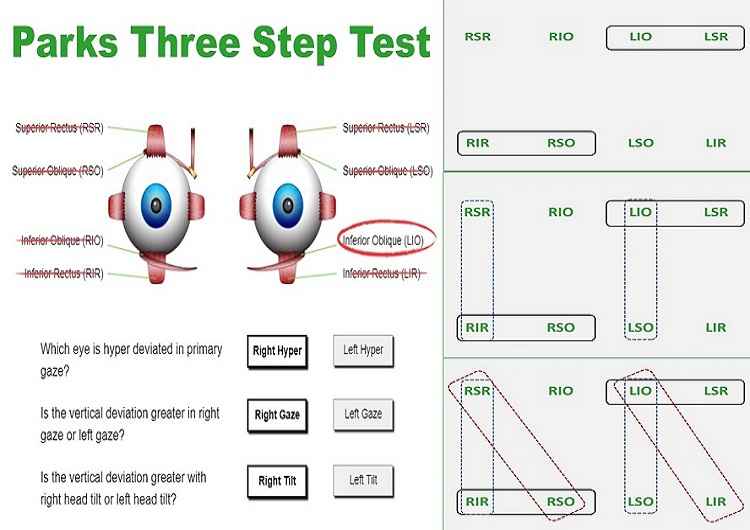
There are in total of eight cyclovertically acting muscles. Four of these muscles work as elevators and four works as depressors of the eyes.
Among these extraocular muscles, the superior oblique and inferior rectus are responsible for the depression of the eyes. Likewise, the inferior oblique and superior rectus elevate the eyes.
At the beginning of the extraocular muscle palsy, there will be limitations in the field of action of the paretic muscle. The overaction in the field of the antagonistic muscle is noted shortly afterward.
Due to the continuous overaction of the antagonistic muscle, there will be a contracture of the antagonist. As a result, there will be a spread of the comitance i.e. the deviation will be equal in all fields of gaze.
At this point, the diagnosis of cyclovertical palsy becomes impossible through the analysis of duction and version movement of the eye. And here comes the important role of the parks 3 step test.
The Parks three-step test can also be utilized to differentiate dissociated vertical deviation (DVD) from other vertical deviations.
Drawbacks
- not reliable for the diagnosis of multiple extraocular muscle paresis or palsy.
- can’t be utilized in case of restrictive strabismus
3 steps in Parks-Bielschowsky head tilt test
Each of the steps in this test eliminates half of the remaining potential extraocular muscles, leaving a single muscle to suspect after the 3 steps.
- The first step is to find out which eye is hypertropic in the primary position.
- The second step in the Parks 3 step test is to determine whether the hypertropia decreases or increases on the right or left gaze.
- The third step is to find out whether the hypertropia increases or decreases upon the right head tilt or left head tilt.
Loose prisms are used to measure the amount of vertical deviation in the primary gaze, side gaze, and in both head tilts. Alternately, a Maddox rod, red filter, and prism can be used together to quantify the vertical tropia of a smaller amount.
How to perform the Parks 3 step test
The procedures of Parks three-step test are performed as follows:
Step 1
The first step is to perform the cover-uncover test in the primary position of gaze and to find out which eye is hypertropic (and hypotropic). If you notice the hypodeviation of one eye then you can consider hyperdeviaiton of the opposite eye.
This step reduces the number of suspected extraocular muscles from eight to four. A right eye hypertropia (or left eye hypotropia) indicates either the weakness of the right eye depressor or the weakness of left eye elevators.
Depressors of the right eye are the right inferior rectus and right superior oblique muscles. Likewise, the left eye elevators are left inferior oblique and left superior rectus.
Similarly, A left eye hypertropia (or right eye hypotropia) indicates either the weakness of right eye elevators or the weakness of left eye depressors.
Depressors of the left eye are the left inferior rectus and left superior oblique muscles. Likewise, the right eye elevators are the right inferior oblique and right superior rectus.
Step 2
The second step is to determine whether the hypertropia is larger in the left or right gaze. Here, the vertical actions of the extraocular muscles are measured. The oblique muscles have their maximum vertical action when the eye is adducted (inward movement), and the recti muscles have their greatest vertical action when the eye is abducted (outward movement).
Step 3
The third step is to find out whether the hypertropia is larger when measured during head tilt to the right or left. During the left head tilt, the left eye intorts and the right eye extorts, similarly the right eye is intorted and the left eye is extorted during head tilt towards the right.
If the hypertropia is larger when the head tilt is to the left, it indicates the weakness of four vertically acting muscles in the left tilt position, and if the hypertropia is larger when the head tilt is to the right, it indicates the weakness of four vertically acting muscles in the right tilt position.
Vertically acting muscles in the right tilt position are RSR, RSO, LIR, and LIO. Similarly, LSR, LSO, RIR, and RIO are vertically acting muscles in the left tilt position.
Caution
- The prism base must be parallel to the floor of the orbit, not parallel to the floor of the room while quantifying the amount of tropia.
- Maddox rod should be held parallel in such a way that the red line is also parallel to the floor of the orbit.
Example of Parks three-step test
Step 1: Find out the hypertropic eye
Let us consider, for instance, the patient being examined has right hypertropia (and left hypotropia), RHT. Draw a solid oval line around the two possible muscle pairs responsible for RHT.

Step 2: hypertropia increases in right or left gaze
If the RHT is larger in the left gaze, it means there is the weakness of any of the following vertically acting muscles in the left gaze: left superior rectus (LSR), left inferior rectus (LIR), right inferior oblique (RIO), or right superior oblique (RSO).
If the RHT is larger in the right gaze, it means there is the weakness of any of the following vertically acting muscles in the right gaze: right superior rectus (RSR), right inferior rectus (RIR), left inferior oblique (LIO), or left superior oblique (LSO).
Suppose the same patient has RHT greater in the right gaze. Draw an oval (dotted lines) around the two possible muscle pairs. So, the suspected muscles are RSR, RIR, LIO, or LSO.
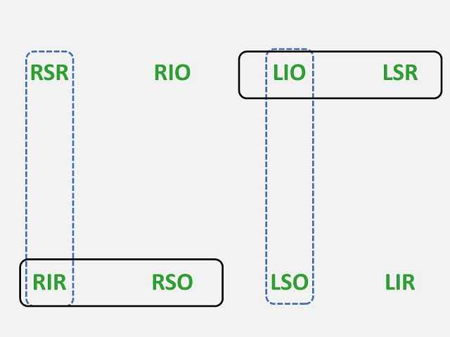
Step 3: hypertropia increases in head tilt toward the right or left
The RHT is quite large when the head is tilted to the right and is almost absent in the head tilt to the left. Again, draw an oval (dotted lines) around the suspected extraocular muscles. So, the suspected muscles are RSR, RSO, LIO, or LIR.

At this point, the LIO is the only extraocular muscle surrounded by three oval lines and it is the affected muscle i.e. the diagnosis is the left inferior oblique palsy.
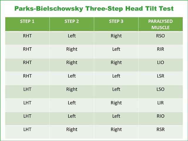
Results of Bielschowsky’s three-step head tilt test in paralysis of all cyclovertically acting muscles are given in the table.
How reliable is Bielschowsky head tilt test
Although the Parks 3 step test is the mainstay for the diagnosis of a cyclovertical muscle paresis or palsy, recent studies have put forward questions on the sensitivity and reliability of this test. In a study “Sensitivity of the three-step test in the diagnosis of superior oblique palsy” by Ajay M Manchandia and Joshep L Demer, they analyzed 50 patients with confirmed evidence of superior oblique atrophy on MRI. They found that the complete three-step test fails to detect 30% of cases of superior oblique atrophy. Often only two of three steps are positive in superior oblique palsy.
In another study “Diagnostic Utility of the Three-Step Test According to the Presence of the Trochlear Nerve in Superior Oblique Palsy”, which was carried out to determine the diagnostic utility of the three-step test in unilateral superior oblique palsy (SOP) according to the presence of the trochlear nerve using high-resolution thin-section magnetic resonance imaging, the results found that the three-step test had a sensitivity of 75% in diagnosing unilateral superior oblique palsy regardless of the presence of the trochlear nerve.
They found the diagnostic utility of the three-step test in unilateral SOP did not differ according to the presence of the trochlear nerve. SR contracture was the main cause of exceptions of the three-step test in the unilateral SOP.
In a study “Errors in the three-step test in the diagnosis of vertical strabismus”, the author mentioned that the use of the three-step test in clinical situations such as contracture of the vertical recti, paresis of more than one vertical muscle, dissociated vertical divergence, previous vertical muscle surgery, skew deviation, myasthenia gravis, and small nonparalytic vertical deviations associated with horizontal strabismus, may lead to incorrect diagnosis and treatment. further imaging tests in addition to the three-step test are necessary to identify a cyclovertical muscle paresis or palsy.
Parks 3 step App and Calculator
Parks 3 step app for iPhone
The Park’s Three-Step Test is used to isolate the paretic muscle in acquired vertical diplopia (vertical tropia). Enter your observations for each of the three tests and the park 3 step app will suggest which extraocular muscle (EOM) is underacting. However, you should never fully trust an app to make your clinical decisions! This app explains how each test is used to determine the final result. Consequently, this application is not only clinically useful but also helps you understand which are the primary EOMs in different fields of gaze.
Download Parks 3 Step App in APP STORE (Paid)
Three-Step Calculator
Enter your observations for each of the three tests and the app will suggest which extraocular muscle (EOM) is underacting. However, you should never fully trust an app to make your clinical decisions! This app explains how each test is used to determine the final result. Visit eYe DOCK for the online calculator of parks 3 step test.
Sources
- A. K. Khurana, Theory and Practice of Squint and Orthoptics (Modern System of Ophthalmology (MSO) Series) 3rd Edition [View]
- Ajay M Manchandia, et al. (2014), Sensitivity of the three-step test in the diagnosis of superior oblique palsy [View]
- Ji Eun Lee, et al. (2017), Diagnostic Utility of the Three-Step Test According to the Presence of the Trochlear Nerve in Superior Oblique Palsy [View]
You May Also Like
Warning signs that you need glasses
ARC Lens or Anti-Reflection Coating Glasses
What Style of Glasses For Face Shape: Oval, Round, & All?
FAQs
1. How is the Parks 3 Step Test performed?
The test has three steps. Step one: With the patient looking straight ahead, cover one eye. Which eye is higher? This tells you which eye has the problem. Step two: Have the patient look left and then right. Is the vertical misalignment worse when looking to the left or to the right? Step three: Have the patient tilt their head to the right shoulder and then to the left shoulder. Is the vertical misalignment worse when tilting right or left? The pattern of answers points to one specific eye muscle.
2. What eye muscles can the Parks 3 Step Test diagnose?
The test diagnoses problems with the four vertical eye muscles. These are the superior rectus, inferior rectus, superior oblique, and inferior oblique. Each muscle moves the eye up or down. A weak or overactive muscle causes double vision. The Parks 3 Step Test can tell you exactly which muscle is the problem. This is important because different muscles need different treatments. Surgery on the wrong muscle will not fix the double vision.
3. Are there apps or calculators for the Parks 3 Step Test?
Yes, several apps and online calculators can help with the Parks 3 Step Test. You enter the results of each step, and the app tells you which muscle is the problem. These tools are great for students learning the test. They are also helpful for busy doctors who want to double-check their work. However, no app replaces a thorough eye exam. The test is just one part of a complete evaluation. Always see an eye doctor for double vision.