All About Rings in Eyes or Rings in Ophthalmology: White, Yellow, Copper, Cholesterol, Blue, and More.
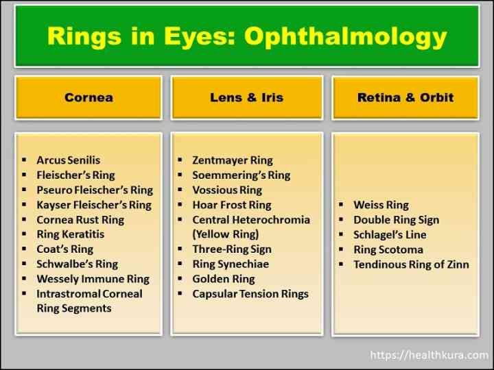
RINGS in EYES or RINGS in OPHTHALMOLOGY gives you a comprehensive list of all rings observed in all structures of the eye; from the anterior-most cornea to the posterior retina. This important list of rings in eyes is equally useful for medical students, medical practitioners, paramedics, nurses, and researchers in the field of medicine.
The list can be a good companion for medical students preparing for any type of entrance exams and university exams. It is a useful nursing guide for those who are participating in USMLE and other academic and professional exams. So, without further ado let’s jump into the topic.

Ophthalmic Rings in Cornea of Eyes

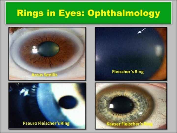
Arcus Senilis (Cholesterol rings in eyes) Blue Rings in Eyes or White Rings in Eyes
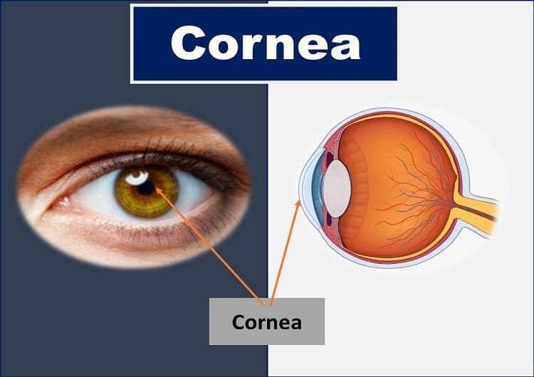
Arcus senilis or the cholesterol ring is a gray, white, or blue ring or arc at the peripheral cornea (anterior-most transparent part) of the eye. It is usually harmless and mostly seen in older adults but can affect people of all ages, and rarely a newborn child.
Cholesterol rings in the eyes can be seen in people below 40 due to high cholesterol levels in the blood. Arcus senilis seen in person under 40 years old is known as arcus juvenilis.
A person with a cholesterol ring or arcus senilis may notice an incomplete, or a complete circle of sharp outer border and blurry inner border on the cornea. As it occurs at the peripheral cornea, the vision remains unaffected in arcus senilis.
Once it appears, arcus senilis will not fade or go away. Some people opt for a risky tattooing process to cover up the cholesterol rings, but it is not recommended by eye doctors. If the cause of arcus senilis is a high cholesterol level in the blood, the doctor will recommend a diet that is low in saturated fats and high in fiber. It is mandatory to quit smoking and increase daily exercise to slow the rate of arcus senilis formation.
Corneal Rust Rings in Eyes
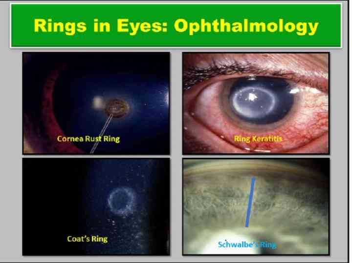
A corneal rust ring is a reddish-brown, circular, or arc structure formed around a foreign body that contains iron and other metals. The rust is formed due to the interaction of metal (iron) with oxygen and other salts of tear just like we see rust with metal left outdoors. The rust formation starts after 3-4 hours of foreign body entry and complete ring formation takes place at around 8 hours.
Corneal foreign bodies and rust rings that are located in the direct axis of vision can cause permanent visual disturbances if improperly removed.
Even after the removal of a foreign body the corneal rust ring may persist but will migrate to the surface of the eye itself within 24 hours. Removal of the corneal rust ring is vital to avoid permanent staining of the cornea or permanent corneal opacity.
The American Academy of Ophthalmology recommends against attempting to remove a residual rust ring at the same time you remove the metallic foreign body due to the possibility of doing further damage to the cornea of the eye.
Rust ring in eye treatment
The eye doctor might use any of the two techniques for the removal of rust rings – hypodermic needle extraction and corneal blurr drill removal. The eye doctor may prescribe prophylactic antibiotic drops or ointment to prevent infection.
In the case of pain, the patient needs to use narcotic pain relievers. Contact lens users should use eyeglasses until complete healing of the eye. Patients should follow up within 24 hours to monitor for proper ocular healing.
Coat’s White Rings in Eyes
Coat’s white ring is a granular, oval ring seen at the level of Bowman’s layer of the cornea due to iron deposition (remnants of a foreign body). It is usually associated with a previous corneal foreign body. Coat’s white rings in eyes are not usually visually significant.
Most of the white rings measure about 0.1 to 0.2 mm in diameter and are usually situated in the periphery of the cornea. George Coats was the first person to describe two cases ‘showing a small superficial opaque white ring in the cornea’(stromal discrete).
Kayser-Fleischer’s Rings: Copper Rings in Eyes
Kayser-Fleischer ring is the most common ocular manifestation of Wilson’s disease. It is also known as the KF ring, Fleischer-Kayser ring, or Fleischer-Strumpell ring. It occurs due to the deposition of copper granules in the Descemet’s membrane of the cornea.
KF ring appears commonly as a golden-brown ring in the periphery of the cornea. It may also appear as greenish yellow, ruby red, bright green, or ultramarine blue. It is almost always bilateral and appears superiorly between 10-2 o’clock first, then inferiorly, and then later becomes circumferential.
Fleischer’s Rings in Ophthalmology
Fleischer’s rings are pigmented rings seen in the peripheral cornea, resulting from iron (in the form of hemosiderin) deposition in basal epithelial cells. Fleischer’s rings are usually seen as a complete or partial ring of yellowish to dark-brown color.
The ring got its name after Bruno Fleischer. Fleischer rings are indicative of keratoconus, a degenerative corneal condition that causes the cornea to thin and change to a conic shape.
Some confusion exists between Fleischer rings and Kayser- Fleischer rings. Kayser-Fleischer rings are caused by copper deposits, and are indicative of Wilson’s disease, whereas Fleischer rings are caused by iron deposits, and are present in Keratoconus.
Pseudo-Fleischer’s ring is observed as a corneal iron deposition in hyperopia.
Schwalbe’s Rings in Ophthalmology
Schwalbe’s line is also known as Schwalbe’s ring in ophthalmology. It is the anatomical line found on the interior surface of the eye’s cornea and delineates the outer limit of the corneal endothelium layer. Schwalbe’s line represents the termination of Descemet’s membrane. This structure is visible only through gonioscopy.

Ring Ulcer or Ring Keratitis or Ring Abscess
Acanthamoeba is a causative organism of ring keratitis. So, it is also known as acanthamoeba keratitis. The vision-threatening, rare parasitic infection was first recognized in 1973. This infection is seen most often in contact lens users.
Stromal ring-shaped infiltrate is the late clinical presentation of ring keratitis. The painful inflammation of the cornea is difficult to diagnose and treat. So, if suspicion of acanthamoeba keratitis arises, the involved region of the cornea is scraped with a sterile instrument (needle, blade, spatula, calcium alginate swab, or cotton tip applicator) under topical anesthesia with the help of a slit lamp.
The culture specimen is then inoculated into a dish of E. coli plated over non-nutrient agar. Acanthamoeba trophozoites and cysts can also be identified with the help of Gram, Giemsa-Wright, hematoxylin, and eosin, periodic acid-Schiff, calcofluor white, or other stains. Confocal microscopy is also used to diagnose Acanthamoeba cysts.
Wessely Immune Rings in Eyes
The Wessely ring or Wessely immune ring is a ring-shaped infiltrate in the corneal stroma that occurs due to a type 3 immune response involving antigen-antibody complex formation. Wessely immune ring may be caused by infections due to acanthamoeba, herpes, pseudomonas, and fungus infection, or sterile etiology.
Wessely immune ring in the eye is seen most often in contact lens users. The painful inflammation of the cornea is difficult to diagnose and treat. Its appearance can aid in the differential diagnosis but can also be misleading. A definitive diagnosis can only be reached when all clinical and microbiological findings are taken into consideration.
In 1911, Wessely first observed reproducible immune reactions in rabbit corneas that had been introduced to foreign horse serum protein and were later challenged with the same antigen. This observation was termed the “Wessely phenomenon” and was further studied many decades later, notably by Morawiecki in 1956, who was the first to observe a ring-shaped infiltrate associated with this reaction.
Intrastromal Corneal Ring Segments (ICRS)
Also known as a corneal implant or corneal insert, intrastromal corneal ring segments (ICRS) are small, crescent-shaped plastic rings that are placed in the cornea to treat keratoconus and pellucid marginal degeneration (PMD). Keratoconus is a worsening disease of the eye in which the normally round cornea bulges into a cone-like shape with an irregular surface, causing distorted vision.
The embedding of the two rings in the cornea is intended to flatten the cornea and change the refraction of light passing through the cornea on its way into the eye.
Rings in Iris and Lens of Eyes

Yellow Rings in Eyes: Central Heterochromia
People with central heterochromia have a different color near the border of their pupils, instead of having one distinct eye color. Yellow rings in the eyes are due to central heterochromia.
The eye may have a shade of gold or yellow around the border of their pupil in the center of their iris, with the rest of their iris another color. It’s this other color that is the person’s true eye color.
Soemmering’s Rings in Ophthalmology
Soammering’s ring is a doughnut-shaped ring or annular swelling of the periphery of the lens capsule. It was first observed by Samuel Sommerring in 1928. He described Soemmering’s ring as deposits of retained equatorial lens epithelial cells that continue to proliferate and form new cortical fibers which eventually form a ring of cortical fibers between the posterior capsule and the edges of the anterior capsule remnant.
Soemmering’s ring forms as a result of anterior capsule edges attachment to the posterior capsule in the absence of intraocular lens implantation or if the lens was not removed in an intracapsular fashion.
It is seen in pseudophakia and also reported in Aphakia or ocular trauma.
Vossius Rings in Eyes
Also known as Vossius’ ring or Vossius’s ring, Vossius ring in the eye is named after German ophthalmologist Adolf Vossius, who first observed and described the ring in 1903. It is seen as a circular ring of fainted or stippled opacity on the anterior surface of the lens due to brown amorphous granules of pigment lying on the capsule after an eye injury.
The ring of pigment is generally the same diameter as the undilated pupil. Diagnosis is made at the slit lamp by observing a complete or incomplete ring of pigment on the anterior lens capsule. The ring is seen most easily with a dilated pupil.
Capsular Tension Rings (CTRs)
Capsular Tension Rings (CTRs) are indicated for the stabilization of the capsular bag in the presence of weakened or compromised zonules. The capsular tension ring maintains the circular expansion and stabilization of the capsular bag.
It is useful in safe intraocular lens (IOL) centration in eyes with zonular dehiscence. It helps prevent IOL decentration after capsular shrinking. Likewise, a capsular tension ring reduces the risk of capsular fibrosis and improves visual acuity when implanted along with premium IOL.
Hoarfrost Rings in Eye
Hoarfrost ring is a feature of pseudoexfoliation syndrome. This ring is formed by the deposition of dandruff-like materials on the lens. It gives a target-like appearance while looking through a slit lamp on the lens.
With an undilated pupil, dandruff-like deposits are seen at the edge of the iris. When the pupil is dilated the appearance is of a granular peripheral zone and central disc on the superficial layers of the capsule, which present with the classical hoarfrost appearance.
Three-ring sign – Rings in Ophthalmology
A three-ring sign is seen on the anterior lens capsule. The ring consists of a central zone of visible exfoliation material measuring 1 to 3 millimeters in diameter, combined with a middle clear zone and a peripheral cloudy ring. The central zone is usually well demarcated and can have curled edges. The middle clear zone is created by the posterior surface of the iris rubbing off the pseudo-exfoliative material from the lens, giving it a target-like appearance.
Ring Synechiae – Rings in Ophthalmology
Ring synechia is also known as annular posterior synechiae. It is formed by the adhesion of the whole rim of the iris to the anterior capsule of the lens. The ring formation prevents the circulation of aqueous humor from the posterior chamber to the anterior chamber, known as seclusion pupillae. As a consequence, the aqueous collects behind the iris and pushes it anteriorly giving rise to “iris-bombe” formation.
The condition in which there is the adhesion of the total posterior surface of the iris to the anterior lens surface is known as the total posterior synechiae.
Zentmayer Ring in Eyes
Zentmayer ring or Scheie stripe, or Scheie line is a circle of pigment seen on the posterior lens capsule at the insertion of the lens zonules which may occur due to pigment deposition in Pigment Dispersion Syndrome (PDS).
In pigment dispersion syndrome, pigment release occurs due to the posterior-bowing of the mid-peripheral iris rubbing against the lens zonules. This unusual iris configuration happens due to a type of “reverse pupil block,” which prevents pressure equalization between the anterior and posterior chambers, leading to a transient rise in the IOP in the anterior chamber relative to the posterior.
Golden Ring in Eyes
A golden ring within the lens of the eye signifies a good, successful hydro delineation during cataract surgery.
YOU MAY ALSO LIKE Lines in Eyes (Ophthalmology): Arlt, Vogt, Haab’s Striae, & All
Ophthalmic Rings in Retina and Orbit of Eyes

Weiss Rings in Eyes
The Weiss ring in ophthalmology is a peripapillary glial tissue that remains attached to the posterior vitreous cortex following posterior vitreous detachment. The ring is composed of fibrous astrocytes and collagen. Patient with Weiss rings commonly gives symptoms of floaters as the ring floats in and out of the visual axis. Posterior eye examination (fundus examination) reveals a partial or complete grey-brownish ring.
The Weiss ring is more common in older people and is most noticeable when moving eyes across a light background. A Weiss ring is a much larger, ring-shaped floater that is created by a posterior vitreous detachment (PVD) from around the optic nerve head. In other words, this is when the vitreous tissue detaches from the retina.
Ring Scotomas – Rings in Ophthalmology
Ring scotoma or tubular vision is the feature of advanced visual field loss in retinitis pigmentosa, glaucoma, and macular diseases.
Roving Ring Scotoma
Roving ring scotoma is noticed in aphakia or high hyperopia correction by eyeglasses (strong plus lens). Prismatic aberration causes roving-ring scotoma or “Jack in the box” effect as a result of which objects keep appearing and disappearing in the field of view.
Flieringa Ring in Eyes
Flieringa Ring or Flieringa Fixation Ring is a stainless-steel ring that is sutured to the sclera to ensure the globe does not collapse during eye surgery. Eight different sizes of the fixation ring are available depending on physician preference and patient variation.
Double Ring Sign – Rings in Ophthalmology
In hypoplasia of the optic disc, the peripheral margin of the encircling ring corresponds to the border of a normal-sized optic disc. This condition is known as a double-ring sign.
The optic disc is often pale or gray, and smaller than normal conditions. A ring of hypopigmentation or hyperpigmentation surrounds the disc defining the area of the putative scleral canal. The outer ring represents the normal junction between the sclera and the lamina cribrosa; the inner ring represents the abnormal extension of the retina and pigment epithelium over the outer portion of the lamina cribrosa.
Tendinous Ring of Zinn – Rings in Ophthalmology
The annulus of Zinn is also known as the annular tendon or common tendinous ring. It is a ring of fibrous tissue surrounding the optic nerve at its entrance at the apex of the orbit. The annulus of Zinn is the common origin of the four extraocular rectus muscles.
The arteries surrounding the optic nerve are sometimes called the “circle of Zinn-Haller” (“CZH”). This vascular structure is also sometimes called the “circle of Zinn”.
The following structures pass through the tendinous ring (superior to inferior):
- Superior division of the oculomotor nerve(CNIII)
- Nasociliary nerve(branch of the ophthalmic nerve)
- Inferior division of the oculomotor nerve(CNIII)
- Abducens nerve(CNVI)
- Optic nerve

Sources
- Isabel W, et. al. Distinctive Wessely immune ring in keratitis-a chameleon, 2020 [Pubmed]
- Campbell DG. Pigmentary dispersion and glaucoma: a new theory. Arch Ophthalmol 1979;97:1667-72.
- http://www.whonamedit.com/synd.cfm/4435.html
- Bhattacharjee H, Deshmukh S (December 2017). “Soemmering’s ring”. Indian Journal of Ophthalmology. 65 (12): 1489. doi:10.4103/ijo.IJO_913_17. PMC 5742989. PMID 29208841. [View]
- Wessely K. about anaphylactic phenomena of the cornea. Munchen Med Wehascher. 1911;58:1713.
- “Soemmering’s ring”. Retrieved 25 March 2018.
YOU MAY ALSO LIKE Wavy Squiggly Lines in Vision (Eye Floaters)

