All About Vogt’s Striae in Keratoconus and Haab’s Striae in Primary Congenital Glaucoma: Introduction, Prevalence, Risk Factors, Diagnosis, Management, Vogt’s Stria vs Haab’s Striae.
Have you heard about two popular striae in the cornea – Haab’s striae and Vogt’s striae? Today, we will be learning in detail about Vogt striae in keratoconus and Haab striae in primary congenital glaucoma. So, let’s get started.

Vogt’s Striae in Keratoconus
Definition of Vogt Striae
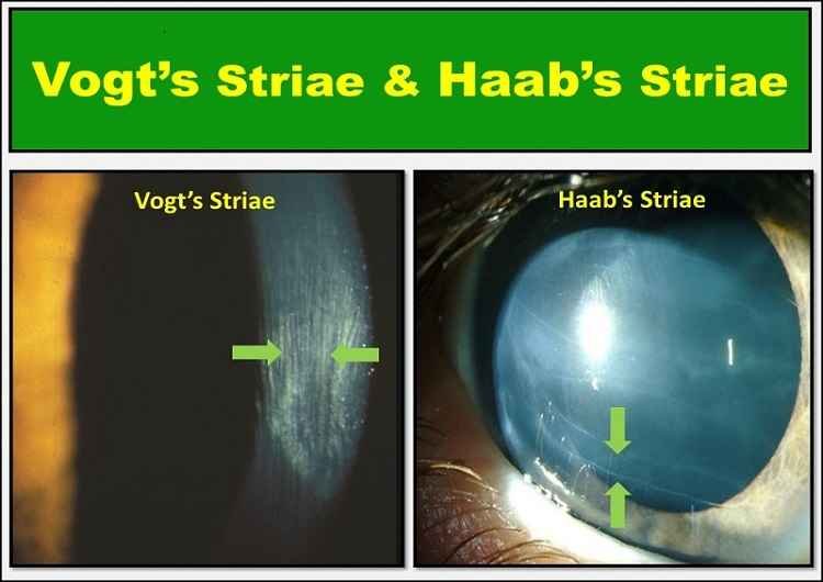
Vogt’s striae are fine vertical (rarely horizontal), whitish lines in the deep stroma, and Descemet’s membrane of the cornea (the anterior-most transparent layer of the eye). Vogt striae are the characteristic features of keratoconus, but can also be seen in the healthy cornea of the eye.
The occurrence of Vogt stria is more frequent in the area of maximum corneal thinning and is best visualized with a wide beam of slit-lamp. The stria can temporarily disappear in the application of external pressure on the globe (at limbus).
The orientation of the lines is related to the steepest axis of the cornea which is formed due to mechanical stress force on collagen lamellae radiating from the cone apex. The striae may be unilateral or bilateral depending on the keratoconus in each eye.
In addition to Vogt’s striae, slit lamp examination can often capture the following common clinical signs in moderate to advanced keratoconus.
- Fleischer’s ring (iron lines partially or completely surrounding the cone)
- Cornea thinning
- Münson’s sign (bulging of the lower lid during downgaze)
- Increased visibility of corneal nerves
- Apical thinning of the cone
- Anterior stromal clearing lines
- Subepithelial fibrillary lines
- Central or eccentric corneal protrusion
- Irregular astigmatism
- Corneal scar
Interesting Fact: Horizontal Vogt’s striae have been reported in only two cases in the literature.

Prevalence and risk factor of Vogt’s striae
According to Zadnik K et al, Vogt striae are found in two-third of the eyes with keratoconus. Similarly, Grieve K et al stated that the Vogt’s striae are seen in 82% of eyes in patients with and without keratoconus.
Eye rubbing is found to be associated with keratoconus formation, with an increased number of striae.
Keratoconus eye (KCN) with Vogt’s striae Versus Keratoconus Eye Without Striae
- KCN eyes with Vogt’s striae have deeper anterior chamber depth and thinner corneas than KCN eyes without striae.
- KCN eyes have worse visual acuity, refractive errors, and corneal tomographic parameters when Vogt’s striae are present.
- Vogt’s striae may be a cause of biomechanical deterioration in keratoconus eyes.
- Vogt’s striae are typically “parallel to the steep axis of the KCN cone.”
- There is a difference in intraocular pressure between eyes with and without striae.
- Eyes with Vogt’s striae have a mean IOP of 13.76 ± 1.16 mmHg, while eyes without Vogt’s striae have a mean IOP of 14.15 ± 1.28 mmHg.
- Vogt’s striae make corneas weaker in KCN eyes. KCN eyes with Vogt’s striae have lower corneal hysteresis (CH), corneal resistance factor (CRH), and Goldman correlated IOP but the greater difference between CH and CRF (CH-CRF) than eyes without striae. Higher CH-CRF indicates worse corneal biomechanics.
- KCN eyes with Vogt’s striae had significantly higher uncorrected and corrected distance visual acuity, maximum keratometry, mean keratometry, and Jackson’s cross-cylinder with axes at 0 and 90 degrees (J0). There was no significant difference in Jackson’s cross-cylinder with axes at 45 and 135 degrees (J45) between the two groups of eyes.
- KCN eyes with Vogt’s striae had lower radius, central corneal thickness, and IOP, but greater deformation amplitude and peak distance.
Diagnosis of Vogt striae
Various ophthalmic instruments that are used to diagnose Vogt’s striae include slit-lamp, optical coherence tomography (OCT), full-field optical coherence microscopy (FFOCM), and confocal microscopy (CM). OCT has the least sensitivity in detecting striae because the width of the striae is at the lateral resolution limit for OCT. Vogt’s striae appear as dark bands on CM, FFOCM, and OCT because the undulated lamellae reflect light away.
Differential diagnosis
Vogt’s striae are similar in appearance to Descemet’s membrane fold. But, it must be differentiated from Descemet’s membrane folds, which can result after surgery to treat keratoconus, such as deep anterior lamellar keratoplasty. Descemet’s membrane folds tend to improve with time and do not have a lasting impact on vision. Descemet folds can also occur due to a pterygium.
Vogt’s striae must also be differentiated from Haab’s striae in congenital glaucoma and Descemet tears due to obstetric trauma.
Management of Vogt striae
If Vogt striae appear in a healthy eye there is no necessity for any treatment. Striae due to keratoconus need to be treated to prevent further deterioration of vision. Treatment of the underlying keratoconus is required to prevent the progression of Vogt’s striae, as KCN eyes with striae have worse biomechanics than KCN eyes without striae.
In healthy eyes, striae may protect against increased IOP, which can be caused by eye rubbing, and do not require treatment.
Haab’s Striae in Primary Congenital Glaucoma
Haab’s Striae Definition
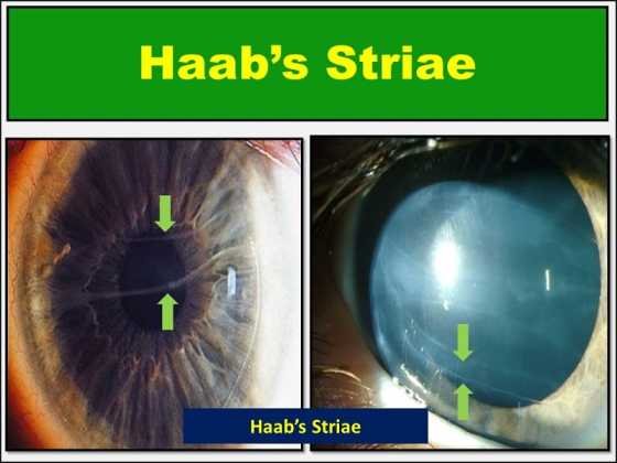
Also known as Descemet’s tears, Haab’s striae are horizontal breaks in the Descemet membrane associated with congenital glaucoma. It is named after Otto Haab (Swiss ophthalmologist). These occur because Descemet’s membrane is less elastic than the corneal stroma.
Tears are usually peripheral, concentric with limbus, and appear as lines with double contours. Vertically oriented breaks in the Descemet’s membrane have been associated with birth trauma (e.g. forceps delivery). Haab’s striae are formed as curvilinear breaks in Descemet’s membrane which occurs due to stretching of the cornea in primary congenital glaucoma.
The orientations of the striae are mostly horizontal, or concentric to the limbus. There may be single or multiple horizontal breaks in Descemet’s membrane (Haab’s stria). Although Haab’s striae are common in infants and children, rare incidences of striae in adults have also been found in the literature. Haab’s striae are the hallmarks of congenital glaucoma.
They are seen as tears in Descemet’s membrane during slit-lamp examination and gonioscopy.

Prevalence of Haab’s Striae
Around one-fourth of the eyes diagnosed with primary infantile glaucoma at birth have Haab’s striae. Likewise, Haab’s striae are seen in more than 60% of those diagnosed with glaucoma at six months of age.
Pathophysiology of Haab’s Striae
Increased intraocular pressure (IOP) leads to expansion of the eye globe. The corneal stroma is more elastic than the Descemet’s membrane. DM is inelastic and tears due to increased IOP. The tears in the DM cause stromal edema, seen in congenital glaucoma.
Tears in the Descemet’s membrane (i.e., Haab’s striae) occur as the cornea stretches under elevated intraocular pressure in primary congenital glaucoma. They are generally visible after the corneal edema had resolved with normalization of the IOP. In specular microscopy, Haab’s striae have been found to be associated with a reduced corneal endothelial cell account.
How Haab’s Striae affect vision?
Haab’s striae can cause astigmatism and affect the clarity of vision when they cross the visual area.
Treatment of Haab’s Striae
Standard glaucoma therapies should be applied early and lifelong monitoring is necessary.
Vogt’s Striae Vs Haab’s Striae
The following table helps to differentiate between Vogt’s striae and Haab’s stria.
| Haab’s Striae | Vogt’s Striae |
| curvilinear or horizontal tears in Descemet’s membrane | vertical (rarely horizontal) folds at the deep stroma or Descemet’s membrane |
| Prevalence: 25-60% of cases in congenital glaucoma | Prevalence: 65% of cases in keratoconus |
| Hallmarks of primary congenital glaucoma | Hallmarks of keratoconus |
| don’t disappear on pressure over the eyeball | disappear with pressure on the eyeball |
| can affect vision due to astigmatism | visually not significant |
Sources
- https://eyewiki.aao.org/Vogt’s_Striae
- https://www.columbiaeye.org/education/digital-reference-of-ophthalmology/glacucoma/optic-nerve-others/haabs-striae
- Güngör IU, Beden U, Sönmez B. Bilateral horizontal Vogt’s striae in keratoconus. Clin Ophthalmol. 2008;2(3):653–655.
- Grieve K, Ghoubay D, Georgeon C, Latour G, Nahas A, Plamann K, Crotti C, Bocheux R, Borderie M, Nguyen T, Andreiuolo F, Schanne-Klein M, and Borderie V. Stromal striae: a new insight into corneal physiology and mechanics. Sci Rep. 2017;7(1):13584
- Bawazeer AM, Hodge WG, Lorimer B. Atopy and keratoconus: a multivariate analysis. Br J Ophthalmol. 2000;84(8):834–836
- Allingham RR, Damiji KF, Freedman S et al. − Congenital Glaucomas. Shield’s Textbook of Glaucoma. Fifth Edition 2005; ch 13: 235-251.
- https://www.atlasophthalmology.net/photo.jsf?node=7349&locale=en
- Wenzel M, Krippendorff U, Unold W et al. − Endothelial cell damage in congenital and juvenile glaucoma.
YOU MAY ALSO LIKE
Diplopia Charting: Common Method of Double Vision Test
Axes of the Eye: Optical, Visual Axis, Angles & More
Hess Charting: Interpretation of Hess Screen Test

