You look in the mirror. One eyelid droops lower than the other. It covers part of your eye. You look tired all the time. You may have trouble seeing. This condition is called ptosis. It is a drooping of the upper eyelid. It can happen in one eye or both eyes.
Ptosis is not just a cosmetic problem. If the eyelid droops enough, it can block your vision. You may have to tilt your head back to see under the lid. Children with ptosis can develop lazy eye. The brain stops using the eye with the droopy lid.
Ptosis can be present at birth. This is congenital ptosis. It is caused by a weak eyelid muscle. It can also develop later in life. This is acquired ptosis. It can be caused by aging, injury, or disease. The most common cause is aging. The muscle that lifts the eyelid stretches and weakens over time.
Treatment depends on the cause and how bad the droop is. For mild cases, no treatment is needed. For more severe cases, surgery can lift the eyelid.
Let’s explore sagging eyelid or droopy eyelid (ptosis), including its causes, treatment, and more.
What is ptosis, droopy eyelid?
Also known as the drooping eyelid, sagging eyelid, blepharoptosis, or ptosis of the eyelid is a condition in which the border of the upper eyelid of one or both eyes falls to a lower position than normal. When a droopy eyelid affects only one eye, it is called unilateral ptosis while in bilateral blepharoptosis both eyes get affected by drooping eyelids.
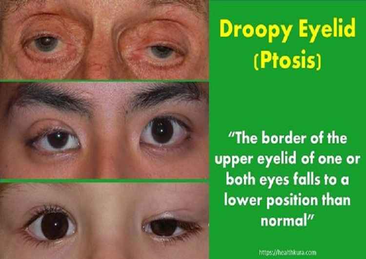
Pathological drooping of the eyelids may occur due to age, trauma, or several medical disorders. Ptosis might be temporary or permanent, and it can be present at birth or can develop later in life. Based on this, blepharoptosis or sagging eyelid is classified as below:
Droopy eyelid: types and ptosis causes
Congenital ptosis of eyelid
Congenital droopy eyelid is associated with congenital maldevelopment of the levator palpebrae superioris (LPS) muscle. It is of four types: simple congenital ptosis, congenital ptosis with associated weakness of superior rectus muscle, blepharophimosis syndrome, and congenital synkinetic ptosis.
A simple congenital sagging eyelid is not associated with any other anomaly. But, blepharophimosis syndrome is comprised of congenital ptosis, blepharophimosis, telecanthus, and epicanthus inversus.
Similarly, congenital synkinetic ptosis is also known as Mascus Gunn jaw winking ptosis and there occurs retraction of the ptotic lid jaw with jaw movements.
Acquired ptosis of eyelid
Acquired ptosis develops later in life. It is classified as neurogenic, myogenic, aponeurotic, and mechanical ptosis.
Neurogenic ptosis occurs with the problem of nerve supply to the LPS muscle. It can be either due to third cranial nerve palsy, Horner’s syndrome, or multiple sclerosis.
Myogenic ptosis or eyelid sagging occurs due to the problem in the LPS muscle itself or in the myoneural junction as in the case of myasthenia gravis, myotonic dystrophy, or trauma to the LPS muscle.
Aponeurotic ptosis happens due to the problem in the connection of LPS to the tarsal plate. It is the most common type of ptosis seen in adults, especially in old age. Clinically the eyelid crease is absent in severe ptosis and often elevated in mild ptosis.
It is the type of acquired ptosis which occurs due to excessive weight on the upper eyelid such as a tumor, edema, dermatochalasis, or anterior orbital lesions.
Pseudoptosis
If there is the appearance of ptosis without levator abnormality, it is called pseudoptosis. The common causes of pseudoptosis are anophthalmos, microphthalmos, enophthalmos, phthisis bulbi, blepharospasm, double elevator palsy, contralateral proptosis, dermatochalasis, and Duane’s syndrome.
Signs and Symptoms of Ptosis or Eyelid Sagging
The main symptom of ptosis is droopy or falling off one or both upper eyelids. It is a painless condition. So, it may go unnoticed in many cases of mild ptosis. The more severe the ptosis gets more it impacts the appearance of the face. Hence, it may cause a psychological impact as well.
You won’t notice any other symptoms if you have mild, uncomplicated ptosis. However, if the ptosis is caused by a serious medical illness, you may find other symptoms related to the underlying cause.
For example, if you have double vision, difficulty speaking, swallowing or breathing, and weakness in the limbs along with droopy eyelid, you may be suffering from myasthenia gravis. Likewise, infants with congenital ptosis might have crossed eyes, lazy eyes, and/or nystagmus.
If ptosis covers part of your pupil and limits the visibility, you might try raising your eyelids unconsciously. As a result, tension-type headaches, and chin up are common in patients with severe ptosis.
Evaluation of Ptosis or Eyelid Sagging
Proper ptosis evaluation is essential to measure the amount of ptosis and can help to successful management of the droopy eyelid. Clinical evaluation of sagging eyelids involves history taking and accurate measurement of margin to reflex distance (MRD), levator function, palpebral fissure height, and superior lid crease.
In history taking, there are five main things to focus on. The onset of ptosis, the family history, history of trauma, history of eye surgery, and variability in the degree of ptosis.
In the clinical evaluation of ptosis, it is necessary to exclude the pseudoptosis and observe if there is a unilateral or bilateral occurrence. Similarly, the function of the orbicularis oculi muscle should be observed carefully as its function is to close the eyelid. The important landmark to notice is the presence or absence of eyelid crease.
It is also necessary to check the jaw-winking phenomenon: the winking motion of the affected eyelid on the movement of the jaw. Another two important things to consider are the associated weakness of any extraocular muscle and the Bell’s phenomenon (an upward and outward movement of the eye while attempting to close the eye).
Now, it’s time to measure the amount of ptosis. The distance between the upper eyelid margin and pupillary light reflex (i.e. marginal reflex distance) is measured using a millimeter scale. It measures how much corneal area is covered by the upper lid. Based on this reading, the severity of ptosis is determined.
- Normal (MRD): less than 2 mm
- Mild ptosis: 2 mm of eyelid droop
- Moderate ptosis: 3-4 mm of droop
Severe ptosis: more than 4 mm of droop (at this stage, the eyelid starts covering the pupil, hence affect vision)
Similarly, the function of LPS should be assessed after the MRD measurement. The examiner holds the patient’s eyebrow with the left hand to prevent the frontalis muscle action. The patient is asked to look down and then to look up. Now, the distance between the lower and upper gaze is measured to determine the ability of LPS to lift the eyelid. The ideal distance should be 12 mm.
- More than 8 mm: good LPS function
- 4-8 mm: moderate function
- Less than 4 mm: very poor LPS function
Droopy Eyelid, Ptosis Treatment (Sagging Eyelid Treatment)
Surgical correction is the mainstay of treatment in congenital ptosis. Surgery should be performed as earlier as possible in severe ptosis to prevent stimulus deprivation amblyopia.
However, surgery can be delayed until the age of 3-5 years in mild to moderate droopy eyelids. The surgical procedures common in ptosis surgery are conjunctive-muller resection, levator resection, and brow suspension.
Plastic surgery is the available surgical option to raise the eyelids in age-related ptosis. This procedure is done under local anesthesia. The surgery is desirable mainly for cosmetic purposes and sometimes to fix visual impairments.
If a person doesn’t want to undergo surgery, special types of glasses are available to correct the droopy eyelid to some extent. The eyeglasses are made with a crutch (ptosis crutch) attachment that can hold up the eyelid.
The ptosis caused by muscle disease, the neurological problems of local eye problems, your doctor will treat the underlying cause which subsequently reduces the effect of a droopy eyelid.
The prognosis of the ptosis surgery is good. In some cases, the eyes may remain open slightly during sleep after surgery. So, it is advised to put nighttime lubricating eye drops or gel to prevent the drying of the eyes.
YOU MAY ALSO LIKE Proptosis or Bulging Eyes
Sources
- What is Ptosis? https://www.aao.org/eye-health/diseases/what-is-ptosis
- Pseudoptosis https://www.aao.org/image/pseudoptosis
FAQs
1. What causes ptosis or droopy eyelid?
Ptosis has many causes. The most common is aging. The levator muscle, which lifts the eyelid, stretches and weakens over time. This is called involutional ptosis. Congenital ptosis is present at birth. It is caused by a weak levator muscle. Injury can damage the muscle or the nerve that controls it. Neurological conditions like myasthenia gravis and Horner syndrome can cause ptosis. So can eye surgery, tumors, and stroke. Sometimes the cause is unknown. See an eye doctor to find out why your eyelid is drooping.
2. How is ptosis treated?
Treatment depends on the cause and how severe the droop is. If the droop is mild and does not affect vision, you may not need treatment. If the droop is caused by a medical condition like myasthenia gravis, treating that condition may fix the ptosis. For most people with age-related ptosis, surgery is the best option. The surgeon tightens the levator muscle so it can lift the eyelid higher. The surgery is usually done as an outpatient. Recovery takes a few weeks. After surgery, your eyelid will look more open and your vision will improve.
3. Can ptosis cause vision problems?
Yes, ptosis can cause vision problems. If the eyelid droops enough, it blocks part of your pupil. This blocks light from entering your eye. Your vision may be blurry or dim. You may have to tilt your head back to see under the lid. This can cause neck pain. In children, ptosis can cause lazy eye (amblyopia). The brain stops using the eye with the droopy lid. The vision in that eye does not develop normally. That is why children with ptosis need treatment early. If you or your child has ptosis, see an eye doctor right away.